
1. Overview
Live blood microscopy provides a close-up view of red blood cells, white blood cells, platelets, and material visible in plasma. The uploaded Howell Medical Supply poster presents numerous examples of microscopic appearances, including separated red blood cells, rouleaux-like formations, cell aggregation, fibrin-like strands, crystals, white blood cells, target-shaped cells, and hemolysis-like changes.
With AI-assisted image processing, LiveBlood.ai is designed to help organize blood microscopy images, recognize visual patterns, and produce consistent educational reports. These capabilities should support, rather than replace, standard laboratory testing and evaluation by qualified healthcare professionals.
2. What Are Live Blood Pictures?
Live blood pictures are microscopic images of a fresh blood sample, commonly viewed using dark-field or phase-contrast illumination. These techniques can make transparent cells and particles appear bright against a dark background.
A conventional clinical blood smear is different. A laboratory professional spreads blood onto a slide, applies a special stain, and examines the size, shape, and number of red blood cells, white blood cells, and platelets. Automated computer programs may also assist with examining clinical blood smears. [medlineplus.gov]
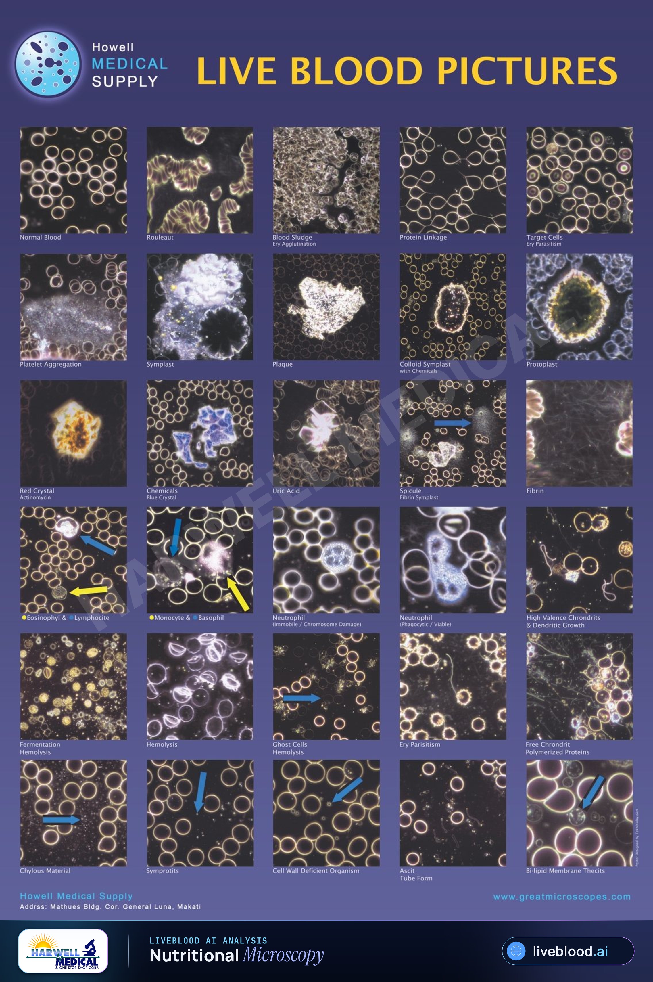
The photographs in the uploaded reference poster show a range of appearances identified by the poster's creator. Several labels represent historical or alternative live-blood terminology and should not be interpreted as confirmed medical findings.
3. Blood Cell Patterns Shown in the Poster
Separated Red Blood Cells
The image labeled "Normal Blood" shows many round cells distributed across a dark background, with visible spacing between a number of the cells.
In a properly prepared conventional blood smear, red blood cells are evaluated for features such as size, shape, color, and distribution. Similar size and color, with a lighter central area, are among the characteristics described for normal red blood cells in standard smear evaluation. [medlineplus.gov]
Microscopic appearance alone, however, does not establish a person's overall health.
Rouleaux-Like Formations
The poster includes an image labeled "Rouleaux," showing red blood cells arranged in curved, stack-like groups.
Rouleaux is a descriptive morphology term. If a laboratory identifies unusual red-cell arrangement, the observation must be interpreted alongside the complete blood count, properly prepared smear, symptoms, medical history, and other relevant tests.
AI image analysis may help flag stack-like arrangements for review, but the software output should not be treated as an independent diagnosis.
Cell Aggregation
The example labeled "Blood Sludge" shows a densely packed field of overlapping and clustered cells. The wording on the poster associates the example with erythrocyte agglutination.
Clustering in a microscopic image can be influenced by medical and nonmedical factors, including specimen preparation, slide pressure, drying, temperature, sample age, and imaging conditions. A qualified laboratory must determine whether a suspected abnormality is genuine or an artifact.
4. Protein Linkage and Fibrin-Like Strands
Several poster images contain thin lines or web-like material between or around cells. The poster labels these patterns as "Protein Linkage," "Fibrin," and "Fibrin Symplast."
The presence of thread-like material in an image does not, by itself, identify its composition or cause. Confirming fibrin or another substance requires an appropriate validated method rather than visual comparison with a reference photograph.
Platelet Aggregation
The poster contains an example labeled "Platelet Aggregation," showing a large granular area surrounded by red blood cells.
Platelets are cell fragments that help blood form clots. A standard blood smear can be used to examine platelets as part of a broader laboratory assessment. [medlineplus.gov]
Possible platelet clumping should be evaluated through proper specimen collection, laboratory procedures, and correlation with a platelet count.
Target-Shaped Cells
One picture is labeled "Target Cells" and shows red blood cells with a ring-like or bull's-eye appearance.
Target cells are recognized in conventional hematology, but their presence is not specific to one condition. MedlinePlus notes that target cells may be associated with abnormal hemoglobin, iron deficiency, liver disease, removal of the spleen, and other factors. [medlineplus.gov]
A healthcare professional must interpret this morphology together with laboratory results and the patient's clinical history.
Crystalline-Looking Material
The poster presents images labeled "Red Crystal," "Chemicals Blue Crystal," and "Uric Acid." These photographs show angular, brightly colored structures among surrounding cells.
Color, brightness, and geometric appearance can vary according to illumination, camera settings, slide preparation, contamination, and optical artifacts. A crystal-like object cannot be reliably identified as a particular chemical or metabolic substance from appearance alone.
White Blood Cells
Several images identify neutrophils, eosinophils, lymphocytes, monocytes, and basophils. These are established categories of white blood cells, which are part of the immune system.
A clinical blood smear may provide the number and types of white blood cells and may reveal abnormal cell shapes. Accurate classification requires proper preparation, staining, laboratory standards, and qualified interpretation. [medlineplus.gov]
Hemolysis and Ghost-Cell Appearances
The lower section of the poster contains examples labeled "Hemolysis," "Ghost Cells Hemolysis," and "Fermentation Hemolysis." These images show pale, distorted, or partially disrupted cell outlines.
Hemolysis means that red blood cells have ruptured and released their contents. Apparent cell damage on a live slide may occur inside the body or during collection, handling, temperature exposure, slide preparation, or prolonged observation. A microscopy image cannot establish the cause without additional evidence.
5. Unverified Historical Terminology
The poster also uses terms such as:
- Symprotits
- Chondrits
- Thecits
- Ascit tube forms
- Symplasts
- Protoplasts
- "Ery parasitism"
- "Cell wall deficient organism"
Some of these terms come from historical or alternative microscopy systems rather than standard hematology terminology. The validity of live blood analysis as a diagnostic laboratory test has not been established, and a Canadian health technology review found limited evidence, with no relevant systematic reviews, randomized trials, or controlled clinical trials identified in its literature search. [cda-amc.ca]
For responsible communication, these labels should be presented as terms printed on the historical reference poster, not as confirmed organisms, diseases, toxins, parasites, or biochemical abnormalities.
6. How LiveBlood.ai Can Support Image Review
Artificial intelligence can support microscopy workflows by analyzing visual properties in uploaded images. Depending on the validated capabilities of the system, useful functions may include:
- Locating visible cells within a microscopic field
- Measuring cell size and shape
- Recognizing clustering or stack-like arrangements
- Highlighting areas for practitioner review
- Comparing images captured at different times
- Organizing image records
- Producing consistent educational reports
AI-generated observations should always remain traceable to the original image. The report should distinguish between a visible feature, a possible interpretation, and a medically confirmed finding.
7. Why Image Quality and Standardization Matter
AI analysis is only as dependable as the images supplied to the system. Differences in specimen collection and image capture may significantly change what appears under the microscope.
Important quality factors include:
- Consistent magnification
- Proper microscope focus
- Stable illumination
- Clean slides and coverslips
- Controlled time between collection and imaging
- Minimal pressure on the blood sample
- Accurate image labeling
- Review of possible collection or preparation artifacts
Standardized imaging helps an AI system compare similar specimens rather than treating technical differences as biological changes.
8. Live Blood Imaging Versus a Clinical Blood Smear
Live blood imaging and clinical blood-smear testing should not be treated as interchangeable.
A clinical blood smear is used with other tests to help diagnose or monitor blood disorders, certain cancers, infections, and parasites. A blood smear alone cannot diagnose a disease. [medlineplus.gov]
LiveBlood.ai should therefore be positioned as an AI-assisted imaging, documentation, and educational platform unless a specific clinical use has undergone the necessary validation and regulatory review.
9. A Responsible Future for AI-Assisted Blood Imaging
The uploaded poster demonstrates the wide variety of structures that may appear in live blood microscopy. It also shows why consistent terminology, careful specimen preparation, and responsible interpretation are essential.
AI may make blood-image review faster and more organized, but trustworthy use requires transparent limitations. LiveBlood.ai can contribute to this approach by helping users document visible patterns without automatically converting every visual feature into a medical diagnosis.
10. Conclusion
Live blood pictures offer an engaging view of red blood cells, white blood cells, platelets, cell arrangements, and material visible in a microscopic field. The Howell Medical Supply poster provides a useful historical and educational collection of these appearances.
LiveBlood.ai brings artificial intelligence into this visual workflow through image organization, pattern recognition, comparative review, and report generation. The most responsible approach is to use these tools for education and practitioner support while referring medical questions to validated laboratory testing and qualified healthcare professionals.
11. Medical Disclaimer
This content is provided for education and general information only. Live blood images and AI-generated observations should not be used independently to diagnose, prevent, or treat a disease. Consult a qualified healthcare professional and use validated laboratory testing for medical concerns.
12. Keep reading
13. Analyze your own blood images
Upload darkfield or brightfield microscopy images and get an 8-section AI report with nutrition guidance and a 7-day meal plan.